












Medical Data Annotation Challenges – and How Healthcare AI Teams Solve Them

Anyone who has spent time on medical annotation projects knows the gap between how it looks on a roadmap and how it actually goes. On paper, annotation is a line item: label the scans, train the model, share with the client. In practice, it’s where a healthcare AI project either earns its accuracy or quietly loses it, scan by scan, long before anyone sees a model metric.
The challenges below aren’t theoretical. They’re the ones that show up repeatedly across imaging modalities, project sizes, and clinical specialties – and they’re the ones healthcare AI companies have learned to work around, rather than wish away.
Challenge #1: The Need for Medical Expertise
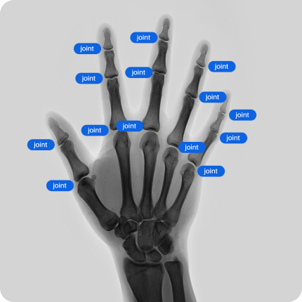
Medical data annotation is not a generic labeling task. A bounding box around a lung nodule or a segmentation mask around a thyroid nodule is only useful if the person drawing it understands what they’re looking at – anatomy, pathology, imaging artifacts, and the clinical reasoning behind why a finding matters.
This is where healthcare AI differs sharply from most other AI domains. A general-purpose annotator can be trained pretty quickly to draw boxes around cars or street signs. Annotating a chest CT for early-stage nodules, or a cranial scan for subtle hemorrhage, requires years of clinical training that can’t be substituted with a style guide, no matter how detailed.

How companies solve it: Most mature annotation programs use a tiered structure rather than assuming every task needs a senior specialist. Non-clinical or junior annotators handle initial passes and repetitive framing tasks, while board-certified radiologists, pathologists, or other domain experts perform review, adjudication, and sign-off on anything clinically ambiguous. This keeps costs manageable without losing clinical accuracy where it counts. The FDA’s guidance on AI/ML-enabled medical devices is explicit on this point: the quality and representativeness of training data is treated as a direct safety factor, not a back-office detail, which is exactly why regulators expect documented clinical involvement in how datasets are built.
Challenge #2: Annotation Variability

Two experienced radiologists can look at the same scan and draw meaningfully different contours around the same lesion. This is known as inter-observer variability, and it isn’t a sign that someone did something wrong – it’s a structural feature of how humans read medical images, especially when a finding sits near a diagnostic threshold.
For AI training, though, variability is a real problem. A model trained on inconsistently labeled data learns inconsistency as if it were a signal. If ten reviewers annotate the same set of scans and disagree on borders, ambiguous findings, or classification thresholds, the resulting dataset teaches the model noise, not truth.
How companies solve it: Structured guidelines are the first line of defense – clear, example-driven annotation protocols that reduce the room for individual interpretation. Beyond that, most serious annotation workflows run inter-observer agreement checks throughout a project, not just as a final QA gate, and use consensus review for cases where annotators disagree. A precision annotation project on pelvic CT scans is a good illustration of this in practice: full-volume bone segmentation required annotators trained on the same tool and workflow, plus a dedicated quality manager whose job was specifically to catch and correct drift between annotators before it accumulated across hundreds of scans.
Challenge #3: Complex 3D Medical Imaging
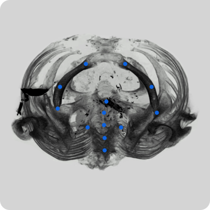
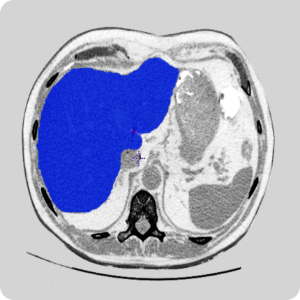
Annotating a 2D X-ray is already demanding. Annotating a volumetric CT or MRI dataset is a different order of complexity entirely. A 3D segmentation task means labeling a structure consistently across every slice of a scan, tracking how it changes shape, and making sure the resulting volume has no gaps, no holes, and smooth, anatomically correct boundaries – all while the underlying anatomy shifts orientation slice by slice.
This is slow, cognitively demanding work, and it’s also where clinical judgment and technical annotation skill have to operate together. A single organ segmentation across a full DICOM volume can take substantially longer than a bounding box task, and the margin for error compounds across every slice.
How companies solve it: 3D annotation workflows lean on specialized tools – platforms like 3D Slicer or Redbrick.AI are common – combined with annotators who’ve been specifically trained on volumetric workflows rather than assuming 2D annotation skills transfer directly. In one breast MRI annotation project, for example, radiologists and trained medical annotators worked from the underlying radiological reports as ground truth, segmenting and labeling each finding to match clinical documentation rather than working from the image alone – a detail worth reading in the full breast MRI case study. That kind of cross-referencing between imaging and clinical notes is often what separates a usable 3D dataset from one that looks complete but doesn’t hold up under model training.
Challenge #4: Ambiguous or Incomplete Labels

Medical findings rarely present as clean, textbook cases. A nodule might sit right at the boundary between two classification categories. An artifact can mimic pathology. A scan can be technically incomplete – missing slices, motion blur, suboptimal contrast – while still containing information a model needs to learn from.
Annotators are regularly asked to make a judgment call where the “correct” label genuinely isn’t obvious, even to an expert. And when annotation guidelines don’t anticipate these edge cases, different annotators resolve the same ambiguity in different ways, which quietly degrades dataset quality in ways that are hard to catch until model performance disappoints.
How companies solve it: The practical fix isn’t trying to eliminate ambiguity – that’s not possible with real clinical data – it’s building a workflow that captures it explicitly. That means annotation guidelines with worked examples of genuinely hard cases, an escalation path for annotators to flag uncertain findings rather than guess, and senior review built in for exactly those flagged cases. Teams that skip this step tend to find out about it only after a model starts producing false positives on the same class of ambiguous case that annotators quietly struggled with during labeling.
Challenge #5: Scalability
A pilot batch of fifty scans, annotated carefully by two specialists, tells you almost nothing about whether that same quality holds at five thousand scans across a dozen annotators. Scaling a medical annotation project isn’t a matter of multiplying headcount – it’s a matter of preserving consistency and clinical accuracy while volume, and usually annotator turnover, increases.
This is where a lot of healthcare AI teams get surprised. The annotation approach that worked cleanly in a small pilot often breaks down once real production volume hits, because the guidelines weren’t stress-tested against the full range of case variety the larger dataset actually contains.
How companies solve it: Scaling successfully tends to follow a specific sequence: validate quality in a pilot phase first, resolve any recurring disagreements or guideline gaps before scaling up, then move to production quickly once quality is proven – rather than letting a long gap open up between pilot approval and full production, which lets institutional knowledge fade. Batch-based delivery, where annotated data is reviewed in chunks rather than all at once at the end, gives teams the chance to catch drift early instead of discovering it across an entire dataset. It’s a workflow discipline as much as a staffing one, and it applies whether the annotation methodology is fast bounding boxes or slow, detailed 3D segmentation.
Challenge #6: Balancing Speed and Accuracy

Every healthcare AI team is under pressure to move fast – investors, product timelines, and competitive markets all push toward shipping sooner. But medical annotation has a floor on how fast it can go without accuracy suffering, and that floor is set by clinical complexity, not by project management preferences.
Push annotators to move faster than the task allows, and errors creep in that are much more expensive to fix later – after a model has already trained on them – than they would have been to catch during labeling. Move too cautiously, and a project loses the timeline advantage that made speed worth prioritizing in the first place.
How companies solve it: The annotation methodology itself is often the lever, not just annotator effort. Bounding boxes are genuinely faster and appropriate for detection-focused tasks where precise contours aren’t clinically necessary; 2D and 3D segmentation are reserved for cases where anatomical precision actually changes the model’s usefulness. Matching the annotation method to what the model genuinely needs – instead of defaulting to the most detailed option everywhere – is one of the more underrated ways teams protect both timeline and quality at once. Realistic per-case time estimates set during a pilot phase, rather than assumed from a spreadsheet, are what make it possible to plan a defensible schedule instead of a hopeful one.
Why This Adds Up to a Data Curation Problem, Not Just a Labeling One
Taken together, these six challenges point at something broader than annotation mechanics: getting medical data model-ready is a curation problem as much as a labeling one. Expertise, consistency, imaging complexity, ambiguity, scale, and speed all interact – solving one in isolation rarely holds up if the others aren’t addressed alongside it. Teams that treat medical data curation and annotation as one continuous discipline, rather than a series of disconnected steps, tend to end up with datasets that actually generalize once a model reaches real clinical settings.
For teams earlier in this process, it’s worth starting with the fundamentals of what medical data annotation actually involves before tackling any of the six challenges above in depth. And for teams about to kick off a new annotation project, the practical sequencing questions – when to pilot, when to scale, how to structure feedback loops – are covered well in a closer look at launching a medical data annotation project.
None of these six challenges have a one-time fix. They’re managed, project after project, through workflow design, tooling choices, and – more than anything else – through annotators and reviewers who understand the clinical stakes behind every label they draw. That combination is what separates a dataset that trains a reliable model from one that just looks finished.
Frequently Asked Questions
What’s the difference between medical data annotation and general-purpose data labeling?
General labeling tasks – tagging a photo as “cat” or drawing a box around a stop sign – can usually be learned by an annotator in an afternoon, with clear right answers most of the time. Medical annotation deals with findings that are genuinely ambiguous even to trained clinicians, requires understanding of anatomy and pathology to interpret correctly, and carries direct consequences for patient safety if the labels are wrong. That’s why medical annotation is typically slower, more expensive per case, and built around clinical review rather than simple task throughput.
Who actually annotates medical imaging data?
It depends on the task’s clinical complexity. Straightforward, well-defined tasks are often handled by trained non-clinical annotators working from detailed guidelines. Anything involving diagnostic judgment – identifying a pathology, contouring a tumor, classifying a finding – is typically annotated or reviewed by radiologists, pathologists, or other relevant specialists, often organized in a tiered structure where junior annotators do the first pass and senior clinicians handle adjudication and sign-off.
How long does it take to annotate a medical imaging dataset?
There’s no single answer – it depends heavily on modality and annotation methods. A bounding box task on an X-ray might take a few minutes per case. A full 3D segmentation of an organ across an entire CT or MRI volume can take considerably longer per case, since the structure has to be labeled consistently across every slice. This is why realistic per-case time estimates, established during a pilot phase rather than assumed upfront, matter so much for planning a project timeline.
What formats and tools are used for medical data annotation?
Most radiology data is stored and annotated in DICOM format, which carries embedded clinical metadata (patient demographics, acquisition parameters, equipment details) alongside the image itself. Other modalities use formats like NIfTI for neuroimaging or whole-slide image formats for digital pathology. Annotation itself is done in specialized tools built for volumetric and clinical data – 3D Slicer and ITK-SNAP are common open-source options, alongside commercial annotation platforms designed specifically for healthcare imaging workflows.
Does more annotated data always mean a better AI model?
Not necessarily. A larger dataset with inconsistent, low-quality, or clinically inaccurate labels can hold a model back more than a smaller, carefully annotated one. This is the practical version of “garbage in, garbage out”: model performance tends to track annotation quality and consistency at least as closely as it tracks raw dataset size, which is why scaling a project has to be paired with scaling quality control, not just annotator headcount.


